Pediatric Heart News
December 13, 2013

“Looking at the pulmonary artery in two dimensions, as we did in the past, you could often miss important obstructions and narrowing,” he says. “I had to take multiple pictures because I knew there was a blockage, but I couldn’t find it from the standard projections.”
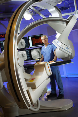
No more. When Johns Hopkins’ Charlotte R. Bloomberg Children’s Center opened in May 2012, it also unveiled a modern pediatric catheterization lab featuring the very latest cardiovascular X-ray biplane imaging designed with three-D capability to improve accuracy and results in procedures like stenting and dilation of narrowed pulmonary vessels.
“You can have the frontal plane swing all around the patient to create a three-dimensional image not unlike that of a CT scan,” says Ringel. “It’s a fantastic option for imaging the child at many different projections and from many different positions to get the necessary information to make the diagnosis.” Also, because the technology has replaced vacuum tubes and image intensifiers with flat panel receptors, Ringel adds, radiation exposure has been reduced by a third.

